|
HOME | SEARCH | ARCHIVE |
|
Miniaturized microscopes
Tiny, user-friendly imaging systems could revolutionize medicine
![]()
By Sarah Yang, Public Affairs
| | 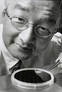 Assistant Professor Luke Lee is developing a micro-lens smaller than the period at the end of this sentence. Peg Skorpinski photo |
13 March 2002
|
Imagine a future where doctors can view the DNA of tumor cells inside a patient as cancer drugs are delivered, or where anti-terrorism units can identify single molecules of a bio-warfare agent on site with a portable detector. With a significant development in miniaturized microscopes, scientists at Berkeley are inching closer to such possibilities. Luke Lee, assistant professor of bioengineering, and his doctoral student Sunghoon Kwon, have captured an image of a plant cell with a micro-lens smaller than the period at the end of this sentence. “It’s shrinking a million-dollar machine down to a size that can balance on the tip of a ballpoint pen,” said Lee, who presented the results at a recent International Conference on Micro Electro Mechanical Systems. “The micro-lens and scanner we’ve made is a crucial part of a microscope that is 500 times smaller than anything in its class.” In testing the accuracy of the micro-lens and scanner, Kwon placed a cell sample taken from a flowering lily, Convallaria majalis, onto the platform of a conventional confocal microscope. Without moving the sample, they captured a cross-sectional image of the cell wall, first with the traditional microscope, then with the micro-lens scanner. They found that the two images matched, showing for the first time that his microscopic lens could perform as well as a conventional one. “Honestly, we were shocked,” said Lee, who also is co-director of the Berkeley Sensor & Actuator Center. “What we’ve finally shown is a proof of concept. We have tested only 2-D images now, but it’s just a matter of time and manpower before we get the first 3-D image.” The micro-lens and scanner are part of a device Lee is developing called the micro-confocal imaging array, or micro-CIA, which in turn is one of a group of devices known as Bio-Polymer-Opto-Electro-Mechani-cal Systems, or BioPOEMS. Invented by Lee, BioPOEMS marry the world of optics to that of microelectromechanical systems, or MEMS, for use in biological applications. The size and sensitivity of the micro-CIA would allow technicians to quickly test even trace amounts of anthrax or smallpox in the field. It could become a crucial part of a “lab-on-a-chip,” where researchers can study genes and proteins in ways unimagined decades ago. The work is being done as part of Berkeley’s Health Sciences Initiative, and Lee is particularly excited by the potential for its advancement in medicine. “You could put this device on the tip of an endoscope that could be guided inside a cancer patient,” said Lee. “Doctors could then see how tumor cells behave in vivo. It would also be feasible to deliver drugs directly to the tumor cell, and then view how the cell responds to the drugs.” High-end confocal microscopes, which house several lasers, take up to a meter of desk space, can cost more than $1 million and typically require highly-trained operators to run them, Lee said. The high cost of owning and running confocal microscopes limits the amount of research that can be done with them. “My goal is to not only shrink the size of these microscopes, but to make them as easy and as cheap to use as a digital camera,” he said.
It is with a hint of populist sentiment that Lee began devising a teeny version of the confocal microscope, the micro-CIA. He envisions a future where confocal microscopy is as common as a Bunsen burner in academic and industry research labs.
Home | Search | Archive | About | Contact | More News
Copyright 2002, The Regents of the University of California.
Produced and maintained by the Office of Public Affairs at UC Berkeley.
Comments? E-mail berkeleyan@pa.urel.berkeley.edu.